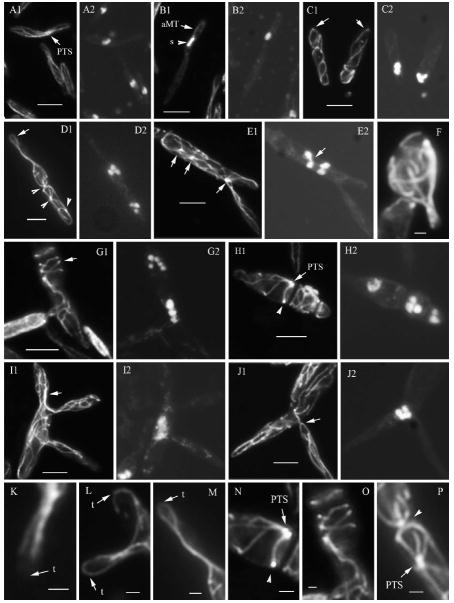
Fig. 9.
Microtubule organization in U. maydis wild type and lis1-depleted cells. Microtubules in wild type and lis1-depleted cells were visualized with an anti-α-tubulin antibody and indirect immunofluorescence. A, B, K. Wild type cells. C–J, L–P. lis1-depleted cells. A1–E1, F, G1–J1, K–P show tubulin staining, and panels A2–E2, G2–J2 the corresponding DAPI-stained cells. K. Microtubules at the cell tip (arrow) of a wild type cell. L, M. Microtubules at the cell tips (arrows) of lis1-depleted cells; M is an enlarged view of the tip of the cell in D1. N, P. Presumed PTS (arrows or arrowheads), N and P are enlarged views of a section of the cells in H1 and D1, respectively. O. An enlarged image of a section of the cell in G1. Bars: A–E, G–J = 5 μm; F, K–P = 1 μm.

