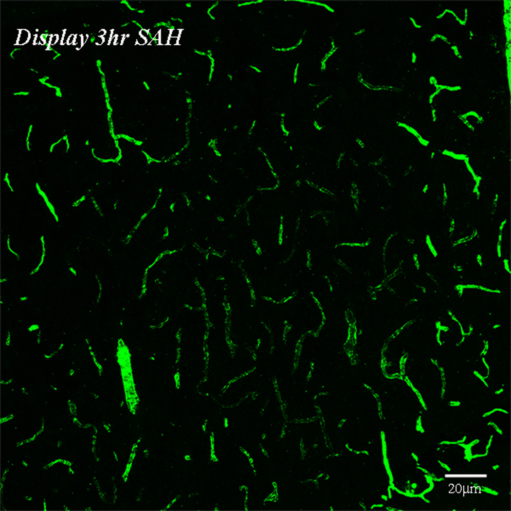
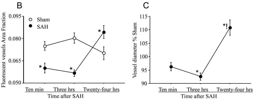
Figure-1. Perfusion deficits in parenchymal vessels after SAH.
Animals were sacrificed at 10 minutes, 3 or 24 hours after Sham or SAH surgeries. Perfusion was visualized as the vascular presence of FITC-dextran injected 10 seconds before sacrifice. A: representative coronal brain sections from animals sacrificed at 3 hours after sham or 3 or 24 hours after SAH surgeries. Note the decrease in FITC-dextran labeled vessels at 3 hours after SAH and an increase at 24 hours as compared to sham. The boxed vessels in the in 3 hour SAH image are displayed in high magnification. B: Temporal change in area fraction of FITC-dextran positive vascular profiles during the first 24 hours after SAH. * significantly different than time matched sham cohorts at P<0.05. C: Temporal change in the diameter of FITC-dextran positive vascular profiles during the first 24 hours after SAH. Note small but significant increase in vessel diameter at 24 hour after SAH. Data are represented as % time matched shams. Mean ± sem, n=5 per time interval per surgery group. *: P<0.05.


