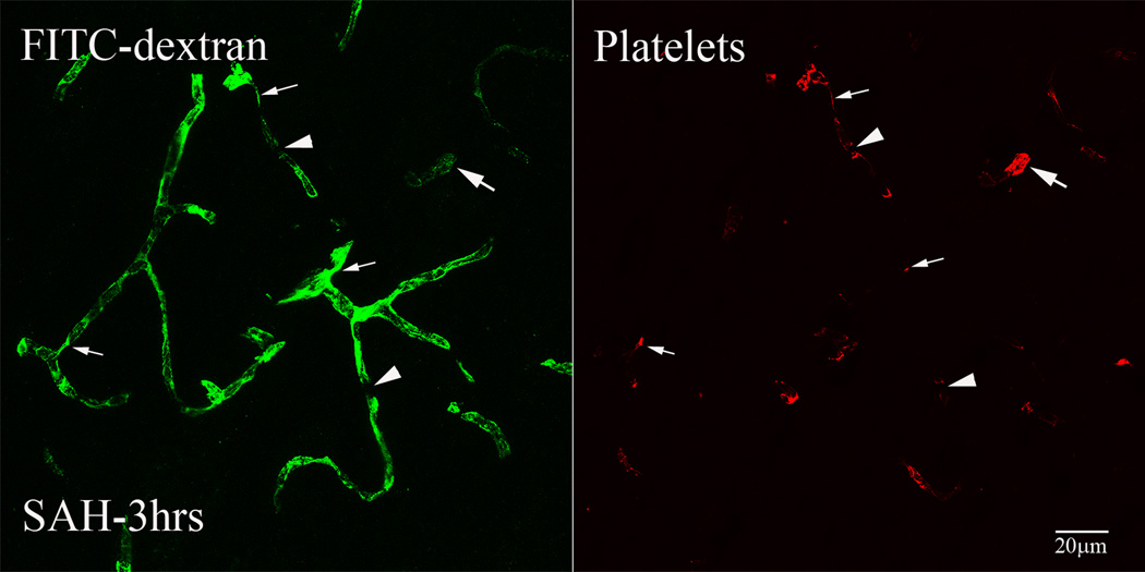
Figure-2. Intraluminal platelet aggregates contribute in perfusion deficits after SAH.
Brain sections from FITC-dextran-injected animals were immunostained for platelets. Shown is a representative 3D rendered stacked high magnification image from animal sacrifice 3 hours after SAH. Two types of vascular segments could be seen; vascular segments filled with platelet aggregates but contain little FITC-dextran label (arrows) and vascular segments with decreased diameter filled with platelet aggregates and FITC-dextran (thin arrows). Some FITC-dextran label vascular profiles appeared interrupted with platelet aggregates present at the interrupted edges (arrow heads).

