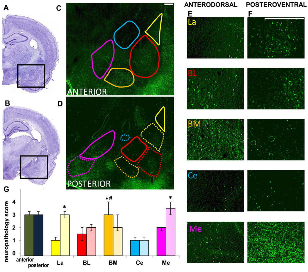
Fig. 4. Neuronal degeneration in amygdalar nuclei, 7 days after soman-induced SE.
A, B. Photomicrographs of panoramic Nissl-stained coronal brain sections showing the anterior (A) and posterior (B) amygdala. C, D. Photomicrographs of Fluoro-Jade C-stained sections of the amygdala, outlining the studied nuclei in the anterior and posterior amygdala (La-yellow; BL-red; BM-orange; Ce-blue; Me-purple). Contours are traced in solid lines for the anterior and dorsal (“anterodorsal”) subdivisions and in dashed lines for the posterior and ventral (“posteroventral”) subdivisions. E, F. Photomicrographs of Fluoro-Jade C-stained sections showing the different anterodorsal and posteroventral amygdala nuclei (scale bar is 300µ for both magnifications). G. Bar graph showing medians and interquartile range of the neuropathology scores for the anterior and posterior sections of the amygdala, and individual amygdala nuclei (solid colors for the anterodorsal and transparent colors for the posteroventral). *p < 0.05 for comparisons between the anterodorsal and posteroventral regions. #p < 0.05 for comparisons between the anterodorsal BM nucleus and the other anterodorsal nuclei.

