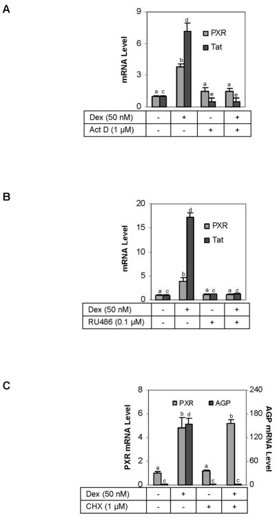
Fig. 3. Effect of actinomycin D, cycloheximide and RU486 on the dexamethasone-induced expression of PXR and Tat (A) Effect of actinomycin D.
H4-II-E-C3 cells were seeded at a density of 5 × 10⁵ in 12-well plates. After an overnight incubation, the cells were treated with dexamethasone (50 nM), actinomycin D (Act D, 1 μM), or both for 6 h. The levels of PXR and Tat mRNA were determined by RT-qPCR. The signals were normalized according to the signal on Pol II mRNA. Letter “b” denotes statistical significance from letter “a”; letter “d” from letter “a” or letter “c”; and letter “a” from letter “c”. (B) Effect of RU486 H4-II-E-C3 cells were seeded as described above and subsequently treated with dexamethasone (50 nM), RU486 (0.1 μM), or both for 24 h. The levels of PXR and Tat mRNA were determined by RT-qPCR, and the signals were normalized according to the signal on Pol II mRNA. Data presented in this figure were assembled from three independent experiments. Letter “b” denotes statistical significance from letter “a”; and letter “d” from letter “c”. (C) Effect of cycloheximide on the dexamethasone-regulated expression of PXR and AGP H4-II-E-C3 cells were seeded as described above and subsequently treated with dexamethasone (50 nM), cycloheximide (CHX, 1 μM), or both for 24 h. The levels of PXR and Tat mRNA were determined by RT-qPCR, and the signals were normalized according to the signal on Pol II mRNA. Data presented in this figure were assembled from three independent experiments. Letter “b” denotes statistical significance from letter “a”; and letter “d” from letter “c”.

