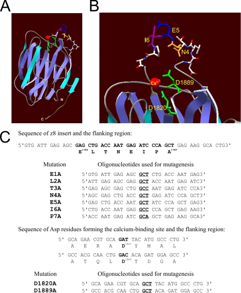
FIGURE 1.
Alanine substitution mutagenesis of AgG3z8. A, β-sandwich folding motif of the calcium-bound AgG3z8 shown in a ribbon diagram. The z8 insert forms a loop between two β-strands on the top edge of the protein (modeled by using laminin globular domain 5 of the laminin α2 chain (αL2LG5; Protein Data Bank code 1DYK, A chain; see supplemental materials). B, enlarged view showing the side chains of the z8 insert and the aspartic acids (Asp1820 and Asp1889) involved in calcium binding. Red, calcium; green, side chains of Asp1820 and Asp1889; orange, side chain of asparagine Asn4. C, synthetic oligonucleotides used for mutagenesis in this study. Only the sense strands of a pair of complementary primers for each mutation are shown. The codons for alanine are shown in bold and underlined. The first and last amino acids of the z8 insert as well as the two calcium binding site aspartates are numbered according to the published rat agrin protein sequence (Protein Identifier G3990241).

