FIGURE 5.
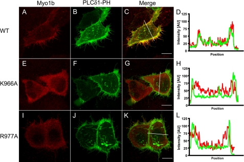
Cotransfection of Myo1b or Myo1b mutants with PLCδ1-PH-GFP in HeLa cells. HeLa cells were transfected with Myc-tagged wild-type Myo1b (A), Myo1b K966A (E) or Myo1b R977A (I) together with PLCδ1-PH-GFP (B, F, and J; green) and then stained with anti-Myc antibody (red). Merged images are shown in C, G, and K for A and B, E and F, and I and J, respectively. D, H, and L show fluorescence intensity along the white lines indicated in C, G, and K, respectively. PLCδ1-PH-GFP, a PIP2-specific binding protein, localized at the cellular periphery and in filopodia (B, F, and J) and colocalized with wild-type Myo1b (C and D). On the other hand, Myo1b K966A (E) did not colocalize with PLCδ1-PH-GFP either at the periphery or in filopodia (F–H). Myo1b R977A did not colocalize with PLCδ1-PH-GFP at the periphery. Scale bars = 10 μm.

