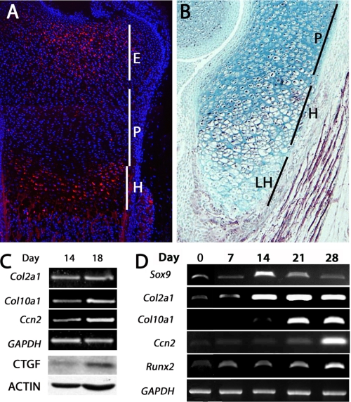
FIGURE 1.
Expression of CCN2 in cartilage. A and B, immunofluorescence and immunohistochemical staining, respectively, of CCN2 expression in E17 (embryonic day 17) (A) and P0 (postnatal day 0) (B) growth plates, show high levels of expression in epiphyseal and hypertrophic chondrocytes, low levels in proliferating chondrocytes, and no expression in terminal hypertrophic cells. C, CCN2 protein and mRNA expression in primary chick chondrocytes is higher in hypertrophic day 18 cells than in proliferating day 14 cells. CTGF, connective tissue growth factor. D, shown is expression of Ccn2 during differentiation of ATDC5 cells. Expression of CCN2 is restricted to hypertrophic stages, which are distinguished from proliferating chondrocytes by expression of Col10a1. E, epiphyseal; H, hypertrophic; LH, late hypertrophic; P, proliferating.

