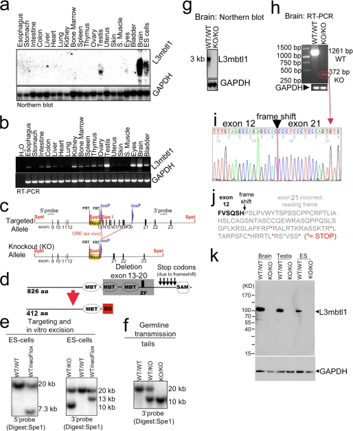
FIGURE 1.
Disruption of L3mbtl1 in mice. a and b, L3mbtl1 expression levels in wild type mice vary among different tissues. S. muscle, skeletal muscle. a, Northern blot analysis demonstrates expression in testes, eyes, and ES cells and most abundantly in the brain. b, expression analysis by RT-PCR reveals widespread, but weaker, expression in other tissues. c and d, strategy for L3mbtl1 disruption (for additional details, see supplemental Fig. S2). d, the knock-out allele lacks the coding region for two MBT domains as well as the zinc finger (amino acids 413 through 708). Additionally, fusion of exon 12 to exon 21 predicts a frameshift that generates several stop codons, so any residual message would not code for the SAM domain but instead express 60 alternative amino acids. e, Southern blot analysis of SpeI-digested DNA from ES cells after targeting and/or Cre-mediated excision of the loxP-flanked regions. As expected, the targeted allele results in a lower band (7.3 kb) compared with the wild type allele (20 kb) using a probe upstream of the targeting vector (5′ probe) (left panel). Correct insertion of the 3′ end was confirmed with a probe downstream of the targeted region (3′ probe; expected bands: wild type, 20 kb; and targeted, 13 kb) (right panel). Excision of the floxed region after transient expression of Cre in vitro results in a 10-kb band (right panel, left lane). f, germ line transmission of the targeted allele documented by Southern blot analysis of tail DNA. g, Northern blot analysis from brains using the entire coding region as a probe demonstrates abundant message in the wild type but no signal in L3mbtl1 knock-out mice. h, RT-PCR analysis for L3mbtl1 using primers upstream and downstream of the deleted region reveals a faint band in the mutant consistent with a shorter message as predicted. i and j, sequence analysis of the PCR product from the mutant in h confirms deletion of exons 13–20 after exon 12 resulting in 60 abnormal residues followed by five stop codons. k, Western blot analysis using an antiserum against a His-tagged fusion protein corresponding to the N-terminal 215 amino acid residues of L3MBTL1 demonstrates absence of the protein in knock-out cells and tissues. Note that a theoretically possible truncated protein (expected size, ∼55 kDa) is not detectable.

