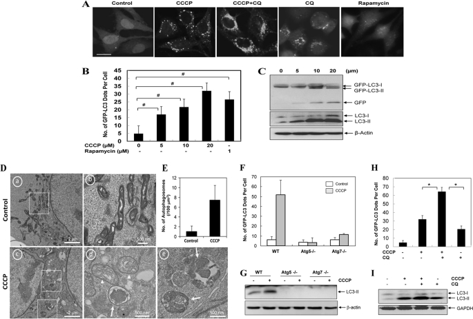
FIGURE 1.
CCCP induces autophagy and mitophagy. A–C, HeLa cells stably expressing GFP-LC3 (GFP-LC3-HeLa) were treated with CCCP at 20 μm (A) or at the indicated doses (B and C) in the presence or absence of CQ (10 μm) for 16 h or with rapamycin (1 μm) for 6 h, followed by confocal microscopy (A). Bar, 20 μm. The number of GFP-LC3/cell was quantified (B), and total lysates were subjected to Western blot analysis with anti-GFP and anti-LC3 (C). D, HCT116 cells were treated with control (panels a and b) or CCCP (20 μm, panels c–e) for 16 h and examined by electron microcopy. Panel b was enlarged from the boxed area in panel a; panels d and e were enlarged from the boxed areas in panel c. E, the number of autophagosomes was counted from more than 12 different cell sections. F and G, wild type (WT), Atg5-deficient, and Atg7-deficient MEF were infected with Ad-GFP-LC3 overnight and then treated with vehicle control or CCCP (30 μm) for 6 h before being analyzed for the number of GFP-LC3 puncta (F) and LC3-II formation (G). H and I, GFP-LC3 HeLa cells were treated with CCCP (30 μm) in the absence or presence of CQ (10 μm) for 16 h. The number of GFP-LC3 dots/cell was quantified (H), and the total lysates were subjected to Western blot analysis with anti-LC3 (I). For B, F, and H, the data shown are the means ± S.D. #, p < 0.05; *, p < 0.01, one-way ANOVA.

