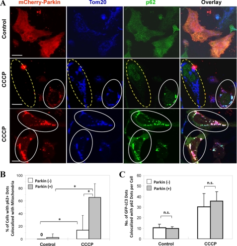
FIGURE 4.
Parkin promotes p62/SQSTM1 mitochondrial targeting following CCCP treatment. A, HeLa cells were transfected with mCherry-Parkin for 24 h and then treated with CCCP (30 μm) for 6 h. The cells were fixed and immunostained for Tom20 and p62/SQSTM1. Bars, 10 μm. The cells marked with yellow dotted ovals had no Parkin expression, whereas cells marked with white solid ovals had Parkin expression and translocation, in which p62/SQSTM1 was also recruited to the mitochondria. B, the numbers of p62/SQSTM1 dots that co-localized with mitochondria per cell were quantified in Parkin-positive and -negative cells. *, p < 0.01, Z test. C, GFP-LC3-HeLa cells were transfected with mCherry-Parkin for 24 h and then treated with CCCP (30 μm) for 6 h. The cells were fixed and stained for p62/SQSTM1. The number of GFP-LC3 dots that were co-localized with p62 were quantified. n.s., no significant difference (p > 0.05): one-way ANOVA. All of the data shown are the means ± S.D.

