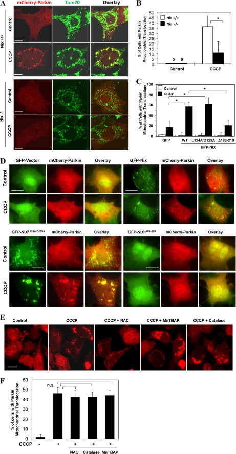
FIGURE 8.
CCCP-induced Parkin translocation is controlled by Nix but not by ROS. A and B, wild type (Nix +/+) and Nix-deficient (Nix −/−) MEFs were transfected with mCherry-Parkin for 24 h and then treated with CCCP (30 μm) for 6 h. The cells were fixed and immunostained for Tom20 (A). Bars, 10 μm. The percentage of cells with Parkin mitochondrial translocation was quantified in Parkin-positive cells. *, p < 0.01, Z test. C and D, Nix−/− MEF cells were co-transfected with GFP vector or GFP-NIX or the mutants, together with mCherry-Parkin for 24 h, and then treated with CCCP (30 μm) for 6 h followed by fluorescence microscopy. Bars, 20 μm. The percentages of cells that had mitochondrial mCherry-Parkin translocation were quantified (means ± S.D.). *, p < 0.01, Z test. E and F, HeLa cells were transfected with mCherry-Parkin for 24 h and then treated with CCCP (20 μm) for 6 h in the presence or absence of NAC (10 mm), MnTBAP (0.5 mm), or catalase (1000 units/ml). The cells were examined by confocal microscopy (C). Bars, 20 μm. The percentage of cells with Parkin translocation was quantified in Parkin-positive cells (D). n.s., no significant difference (p > 0.05), one-way ANOVA. All of the data shown are the means ± S.D.

