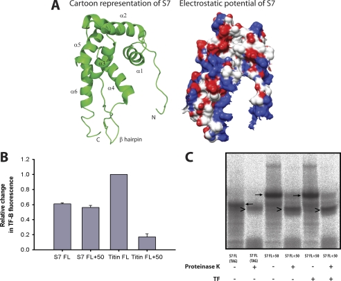
FIGURE 7.
Interaction of TF with RNCs of the ribosomal protein S7. A, ribbon diagram and electrostatic surface potential of the crystal structure of S7 from the intact E. coli ribosome and shown in the same orientation (PDB code 2AVY). Surfaces are shown in degrees of positive (blue) and negative (red) potential. Electrostatic potential was generated with Chimera (50). B, the maximal relative change in TF-B fluorescence, reflecting TF recruitment to the ribosome, measured with nascent chains of S7 FL, S7 FL+50, Titin FL, and Titin FL+50. Values were corrected for the amount of radiolabeled protein synthesized. The fluorescence change of TF-B with Titin FL is set to 1. C, proteinase K digestion of S7 FL (TAG) (ribosome released S7) or S7 FL+50 nascent chains. The nascent chains were translated in the PURE system for ∼50 min in the presence of [35S]methionine. S7 FL+50 nascent chains were translated in the presence or absence of 5 μm TF. The reactions were digested with Proteinase K for 8 min on ice and analyzed by SDS-PAGE. Black arrows indicate nascent chains, arrowheads indicate protease-protected fragments.

