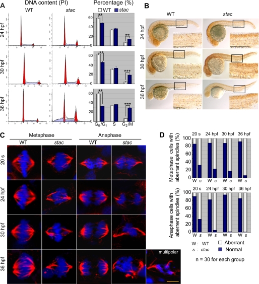
FIGURE 6.
stac mutant embryos have more abnormal mitotic cells. A, cell cycle analysis by flow cytometry. Cells from stac mutant or wild type (WT) sibling embryos at indicated stages were labeled with propidium iodide (PI), and DNA content was counted. The percentages of cells in different phases of the cell cycle are shown in bar graphs. Significance of differences is as follows: **, p < 0.01; ***, p < 0.001 (by Student's t test). Error bars indicate S.D. B, mitotic cells labeled with anti-pH3 antibody in stac mutant and wild type sibling embryos at indicated stages. The boxed area is enlarged at the right corner of each panel. Note that more mitotic cells are found in stac mutants. C and D, mitotic spindles and chromosome aggregation and segregation at metaphase and anaphase of mitotic cells in stac mutant and wild type sibling embryos. Immunohistochemistry was performed with anti-α-tubulin for spindles (red) and DAPI for chromosomes (blue) (C). Cells were picked up from the spinal cord. An abnormal mitotic cell in the anaphase from a mutant embryo was shown to have multipolar spindles (on the right lower corner of panel). Scale bar, 5 μm. The percentage of mitotic cells with aberrant spindles is shown in D.

