FIGURE 1.
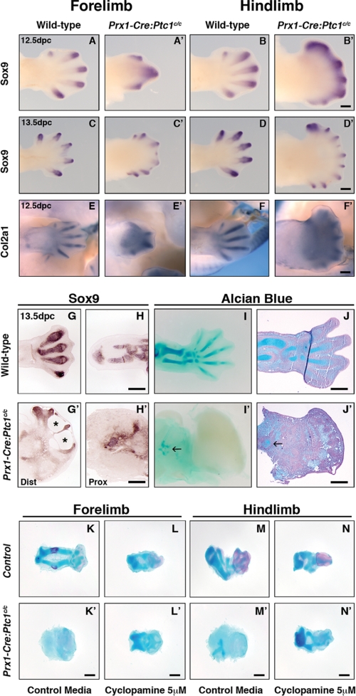
Altered chondrogenic gene expression and structural abnormalities in Prx1-Cre:Ptc1c/c limbs. Compared to wild type (A, B, C, and D), whole mount in situ hybridization for Sox9 revealed expression at distal tips of Prx1-Cre:Ptc1c/c digits at 12.5 dpc (A′ and B′) and 13.5 dpc (C′ and D′), along with ectopic inter-digital expression at 12.5 dpc. Localized expression of Col2a1 was also disrupted in the Prx1-Cre:Ptc1c/c limb at 12.5 dpc (E–F′). Scale bar, 500 μm. At 13.5 dpc, section in situ hybridization for Sox9 revealed disorganized and reduced expression in the distal autopod (dist) and proximal zeugopod (prox) of the limb (G, G′, H, and H′). Whole mount (I and I′) and section (J and J′) Alcian blue staining confirmed disrupted cartilage development. Arrows in I′ and J′ show small regions of Alcian blue staining in mutant limbs. Scale bar, 200 μm. Inhibition of hedgehog signaling with cyclopamine rescues the chondrogenic defect in Prx1-Cre:Ptc1c/c limbs cultured ex vivo. Whole autopods were harvested at 11.5 dpc and cultured for 6 days. Addition of cyclopamine perturbed Alcian blue staining in control limbs (K–N), but partially rescued the phenotype in limbs derived from Prx1-Cre:Ptc1c/c embryos (K′–N′). Images are representative of four individual experiments (see supplemental Figs. 2 and 3). Scale bar, 200 μm.

