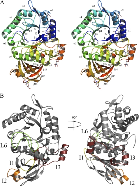
FIGURE 2.
Structure of ScAGal monomer. A, schematic stereo representation of the ScAGal monomer showing a catalytic (α/β)8 domain and a β-sandwich domain. Secondary elements from both domains are labeled. Glycosylated Asn and GalNAc moieties are shown as sticks. B, two views of ScAGal monomer highlighting the important insertions in loops L6 (green), I1 (yellow), I2 (orange), and I3 (maroon).

