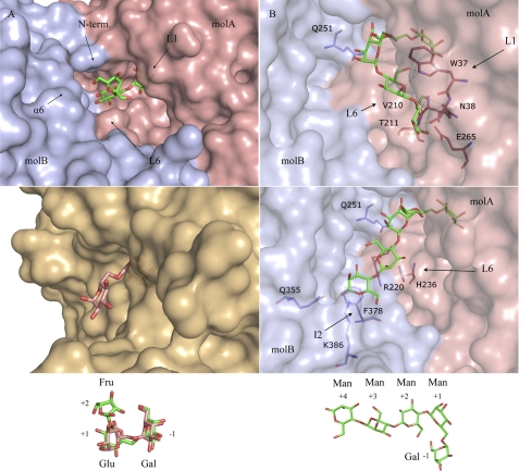
FIGURE 6.
ScAGal active-site specificity. A, comparison of the ScAGal-raffinose (top) and human α-galactosidase-melibiose (bottom, Protein Data Bank code 3HG3) complexes, showing the deep cavity that give access to the ScAGal catalytic pocket. The scheme shows the fixed conformation of raffinose, which is rotated by 180° around the −1/+1 glycosidic bond with respect to the melibiose found in the human enzyme. B, two putative positions of an α-1,6-galactose-substituted manno-tetrasaccharide (shown in the scheme), docked into the ScAGal active-site as explained in the text. The residues that may be involved in shaping subsites +1 to +4 are highlighted.

