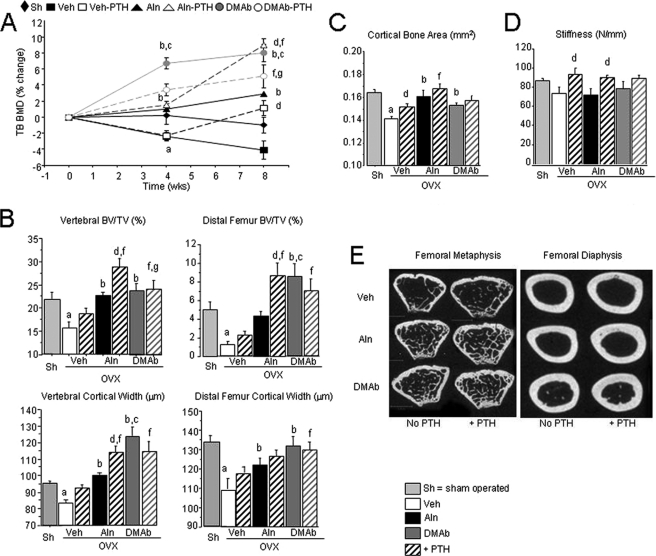
FIGURE 2.
BMD, bone microarchitecture, and strength in OVX huRANKL mice treated with Aln or DMAb, with or without PTH. A, effects of the different noted therapies on TB BMD. Data are expressed as the percentage change from baseline (0 week), DXA (± S.E.). Vertebral and femoral cancellous (B) and cortical (C) microarchitecture at the midshaft femur, and femoral stiffness (D) in OVX huRANKL mice treated with Aln or DMAb, with or without PTH, for 8 weeks post-OVX. E, two dimensional micro-CT representative images of the femoral metaphysis and diaphysis for each treatment group. a, p < 0.05 compared with Sham; b, p < 0.05 compared with OVX Veh; c, p < 0.05 compared with OVX Aln; d, p < 0.05 compared with non-PTH in the same pretreatment group; e, p < 0.05 compared with baseline; f, p < 0.05 compared with OVX Veh-PTH; g, p < 0.05 compared with OVX Aln-PTH.

