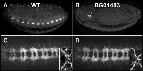
Figure 6.
Normal CNS development in the absence of Hsp23. (A, B) Ventro-lateral view of Hsp23 expression at stage 16. (A) Wild-type embryo. (B) Embryo homozygous for the P-element insertion BG01483. Note the absence of Hsp23 expression in the CNS. The Hsp23-positive cells located behind the embryonic brain are tentatively identified as the crystal cells of the immune system. (C, D) Ventral views of CNS ultra structure in stage 16 embryos using the BP102 antibody. Insets show the anterior VUMs axonal projections visualized with the 22C10 antibody. (C) Wild-type embryo. (D) Embryo homozygous for the P-element insertion BG01483. No differences are detected between C and D.

