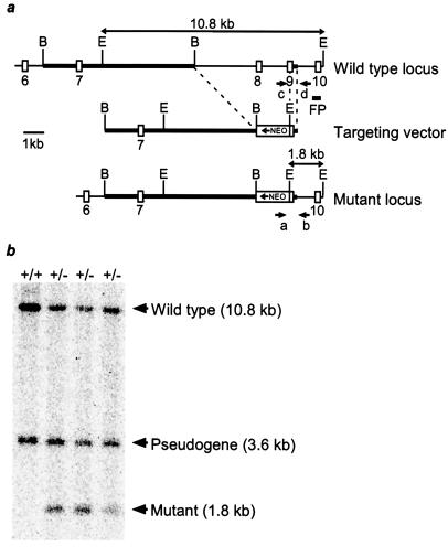
Figure 1.
Targeted disruption of the murine tsg101 locus. (a) Partial genomic organization of the mouse tsg101 locus (Top) and structure of the targeting vector (Middle). Coding exons are depicted as open boxes and homologous regions in the targeting vector are indicated by thickened lines. In the mutant allele (Bottom), a PGKneo cassette replaced 4.4 kb of the tsg101 locus, encompassing exon 8 and most of exon 9, in opposite orientation to tsg101 gene transcription. Positions of the PCR primers (a–d), 3′ flanking probe (FP) used in Southern blot analysis, and predicted sizes of restriction fragments for genotyping are shown. B, BamHI; E, EcoRI. (b) Southern blot analysis of ES cell clones generated by homologous recombination at the tsg101 locus. Genomic DNA from wild-type (+/+) and heterozygous ES cell clones (+/−) was digested with EcoRI and hybridized to the indicated 3′ flanking probe. The 1.8-kb fragment is diagnostic of the mutant allele.

