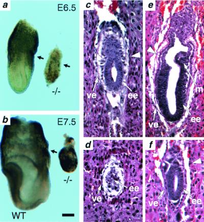
Figure 2.
Severe developmental delay in tsg101 mutant embryos. Morphology and histological analysis. (a) E6.5 tsg101−/− embryos (−/−) are smaller and less organized than their wild-type littermates (WT). The arrows point to the separation between the embryonic and extraembryonic regions. (b) E7.5 tsg101−/− embryos have failed to progress and are starting to be resorbed. (c) E5.5 wild-type embryo at the early egg cylinder stage. (d) E5.5 tsg101−/− embryo showing a poorly defined extraembryonic region and disorganized visceral endoderm. (e) E6.5 wild-type egg cylinder stage embryo. Both the embryonic and extraembryonic regions are well-organized and nascent mesoderm tissue can be distinguished. (f) E6.5 tsg101 mutant embryo. The embryonic and extraembryonic regions are severely underdeveloped and no mesoderm is observed. The large arrowhead in c, e, and f points to the separation between the embryonic and extraembryonic regions. ee, embryonic ectoderm; ve, visceral endoderm; m, mesoderm. (Bar = 60 μm in a, c–f; 120 μm in b.)

