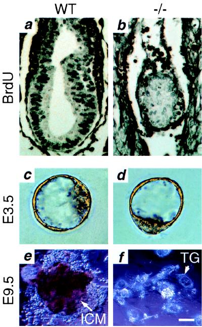
Figure 4.
Reduced cellular proliferation in tsg101−/− embryos in vivo and in vitro. (a and b) BrdUrd incorporation. (a) Strongly BrdUrd-positive nuclei can be seen throughout an E6.5 wild-type embryo. (b) Fewer BrdUrd-positive cells with a much weaker signal are seen in an E6.5 tsg101−/− embryo. (c—f) ICM outgrowth. (c) Wild-type E3.5 blastocyst. (d) Tsg101−/− E3.5 blastocyst. (e) Wild-type outgrowth after 6 days of culture. The ICM is surrounded by trophoblast giant cells (TG). (f) Outgrowth of tsg101−/− blastocysts after 6 days of culture. Only TG cells remain. (Bar = 80 μm in a and b; 20 μm in c and d; 40 μm in e and f.)

