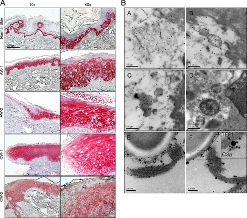
FIGURE 7.
Identification of TFPI in human skin and wounds. A, immunohistochemical identification of TFPI in normal skin, acute wound (AW), and in chronic venous leg ulcer tissue (chronic wounds, CW). Skin biopsies were taken from normal skin (n = 3), from acute wounds (AW-1 and AW-2, 5 and 8 days after wounding, respectively), and from the wound edges of patients with chronic venous ulcers (CW, n = 3). Representative sections are shown. In normal skin TFPI is detected mainly in the basal layers of epidermis. TFPI in AW and CW are ubiquitously found in all epidermal layers. Scale bar, 100 μm. B, TFPI-derived peptides are found in human wounds. Visualization of binding of C-terminal TFPI peptides to bacteria found in fibrin slough from a P. aeruginosa-infected chronic wound surface. The peptides bind to fibrin (A), bacteria (B and C), and bacteria inside a macrophage (D). In E and F, TFPI and C3a peptides were visualized by immunogold using gold-labeled antibodies of different sizes, specific for C3a (10 nm) and C termini of TFPI (20 nm), respectively. Evaluation of 50 bacterial profiles showed that ∼70% of TFPI-molecules were associated with C3a. See inset for exemplification.

