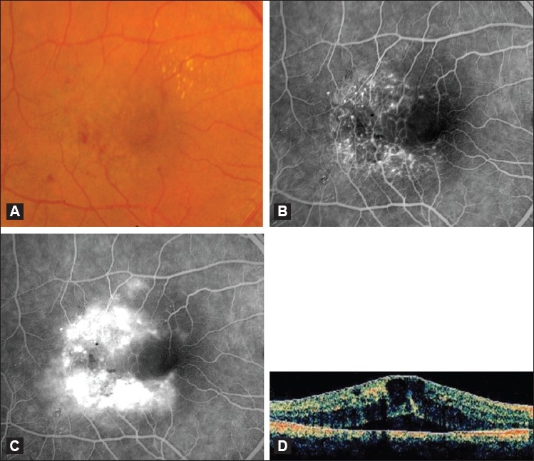
Figure 1.

(A) Color fundus photograph of an eye with IJFT I. Note the visible retinal telangiectasis, aneurysmal dilations temporal to the fovea, and the characteristic lipid deposition. (B and C) Corresponding early (B) and late (C) fluorescein angiogram showing easily visible telangiectatic vessels and aneurysmal capillary dilations straddling the horizontal raphe causing late leakage. (D) Optical coherence tomography scan of an eye with IJFT I showing increased central retinal thickness, intraretinal fluid-filled spaces, and subretinal fluid
