
Figure 5.
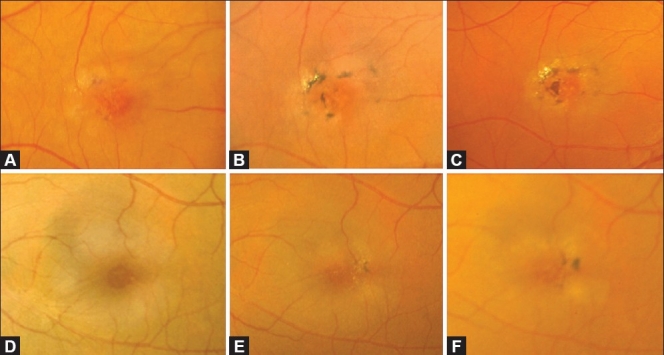
Serial color fundus pictures of the right eye (A-C) and left eye (D-F) of a patient with JFT IIA. (A) Early Stage 4 depicting intraretinal pigment epithelial deposition in the vicinity of the temporal right-angled venule. (B and C) Over a period of 5 years, a progressive increase in the number and size of the intraretinal pigment clumps was observed. The clumps are located preferentially close to the nearby parafoveal vessels. Note the increasing superficial refractile crystals over time, and the central foveal atrophy which is more clearly seen in (C). In the left eye, which was at an earlier stage (Stage 2 in D) similarly showed increasing intraretinal pigment epithlelial migration over time (Stage 4 in E and F). This example illustrates that IJFT II can be asymmetric. Note how the foveal depression simulates a macular hole in this eye (D)