
Figure 8.
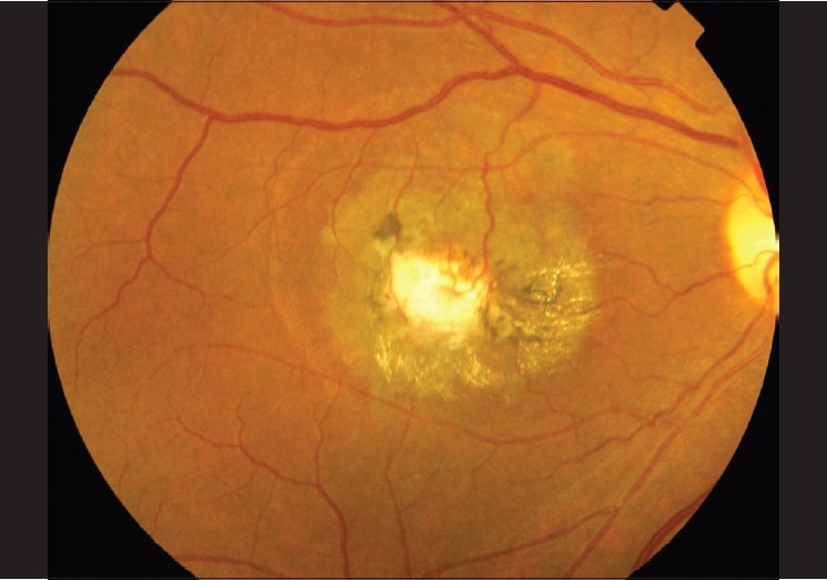
Color fundus photograph of IJFT IIA late Stage 5 that did not receive treatment showing fibrovascular tissue. Note the retinal venules dipping into the fibrous tissue. The superficial retinal crystals are still present
Official websites use .gov
A
.gov website belongs to an official
government organization in the United States.
Secure .gov websites use HTTPS
A lock (
) or https:// means you've safely
connected to the .gov website. Share sensitive
information only on official, secure websites.
Color fundus photograph of IJFT IIA late Stage 5 that did not receive treatment showing fibrovascular tissue. Note the retinal venules dipping into the fibrous tissue. The superficial retinal crystals are still present