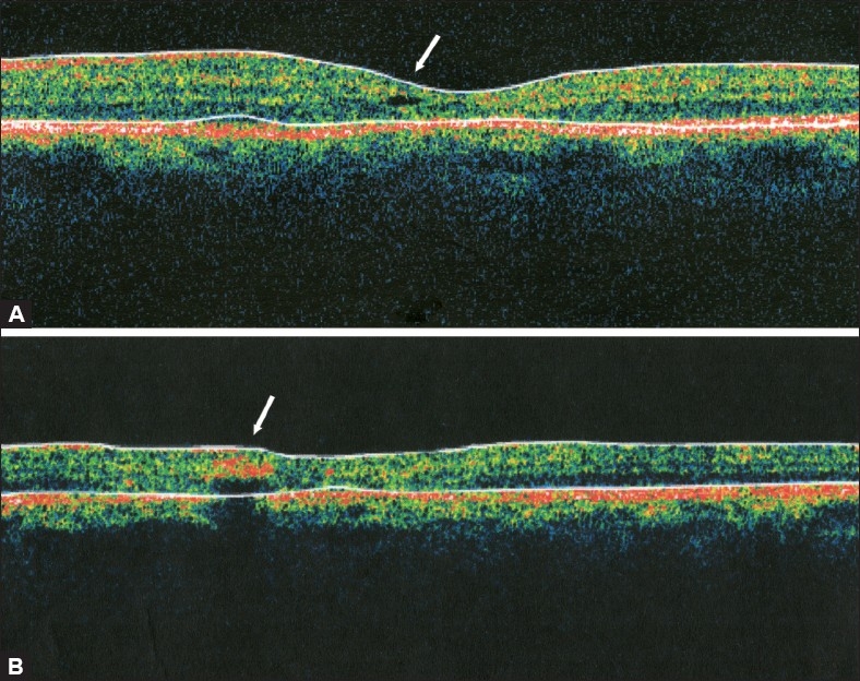
Figure 9.

(A) Optical coherence tomogrpahy scan at the fovea demonstrating a small inner lamellar retinal cyst “cystoid” (arrow) typical of IJFT IIA. Note the absence of retinal thickening or fluid-filled spaces. (B) Optical coherence tomogrpahy scan demonstrating an intraretinal hyper-reflective area (arrow) that corresponds to an intraretinal pigment epithelial plaque which causes posterior shadowing
