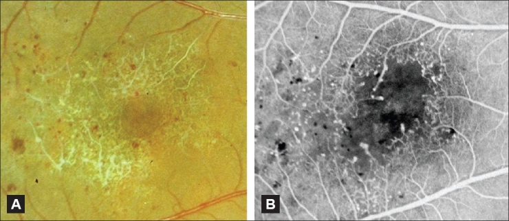
Figure 10.

(A and B) Color fundus photograph (A) and corresponding fluorescein angiogram (B) of an eye with occlusive IJFT (group III). Note the marked perifoveolar capillary nonperfusion seen clinically and angiographically. There are numerous telangiectatic vessels as well that cause limited exudation along the vascular walls. Limited exudation in IJFT III was described by Gass and Blodi[2]
