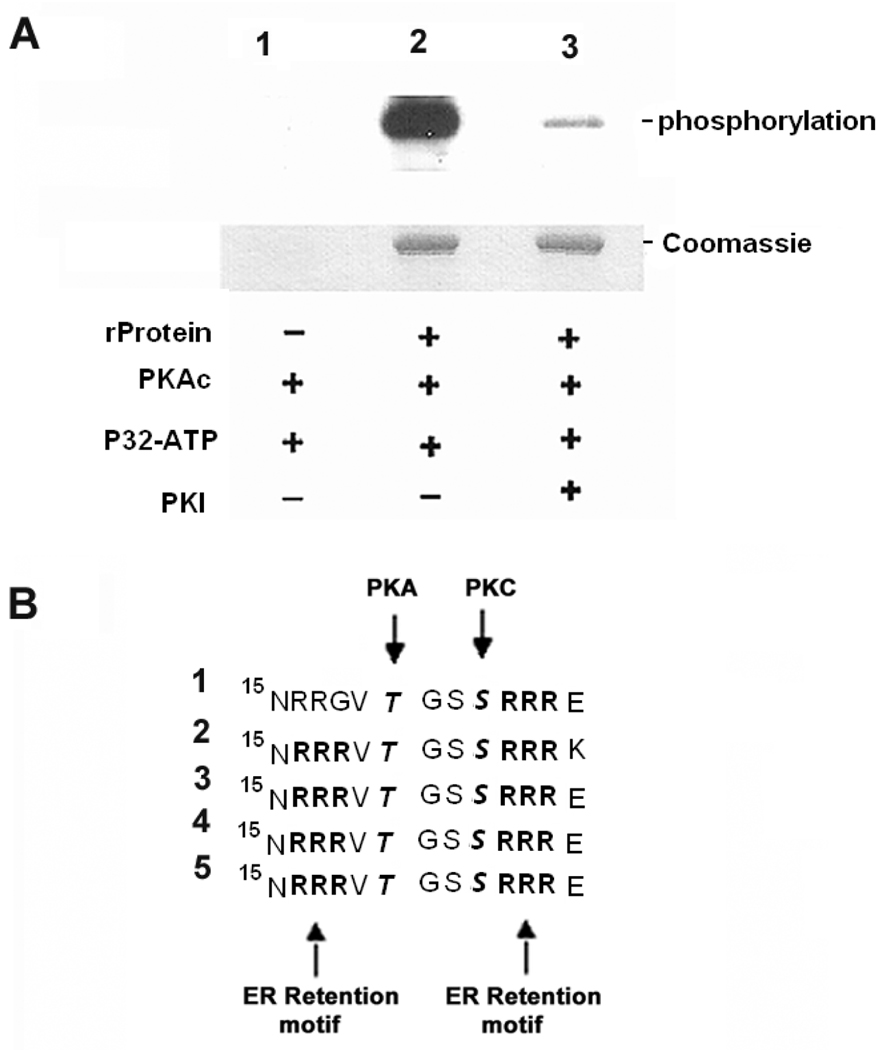
Fig. 3. PKA phosphorylates trans-sialidase recombinant protein in vitro PKAc phosphorylation sites were found to flank ER retention motifs in the members of trans-sialidase superfamily.
A. In vitro phosphorylation of trans-sialidase recombinant protein by PKA is confirmed.
Lane1, Absence of trans-sialidase recombinant protein as negative control;
Lane 2, Trans-sialidase recombinant protein was phosphorylated in absence of PKI;
Lane 3, PKI inhibited the phosphorylation; − absence of the reagent; + presence of the reagent (for details see MATERIALS AND METHODS).
B. Five trans-sialidase protein sequences interacting with TcPKAc possess RXR (Arg-X-Arg) ER retention motifs in N-terminal. A PKAc phosphorylation site and a PKC phosphorylation site flank with these sites. It has been reported that phosphorylation by PKA and PKC in ER and /or Golgi at these sites suppresses ER retention and regulates surface delivery to membrane. The sequences were lined up at N-terminal position 15, RXR is a typical ER retention signal; RRXT or RXXT (position17–20) is a PKA phosphorylation site and SXR (position 23–25) is a PKC phosphorylation site.
(1). Trans-sialidase Tc00.1047053508563.20;
(2). Trans-sialidase Tc00.1047053509187.10;
(3). Trans-sialidase Tc00.1047053507839.40;
(4). Trans-sialidase Tc00.104753510111.20;
(5). Trans-sialidase Tc00.1047053508285.60.

