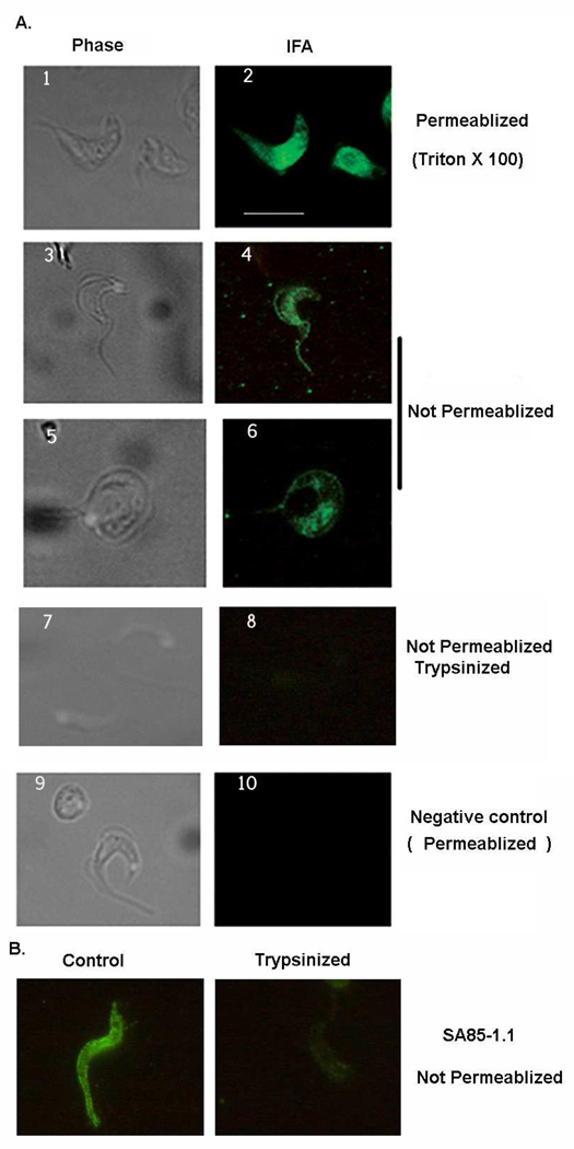
Fig. 5. TcPKAc localizes both in cytoplasm and on membrane.
A. IFA was performed in trypomastigotes. With treatment of Triton X-100 for permeabilization, TcPKAc staining is evident in the cytoplasm (1–2 panel). Without permeabilization (no Triton X-100), the antibody also recognized TcPKAc on the surface membrane and flagellum (3–6 panels). Trypsin treatment dramatically reduced surface staining with TcPKAc mAb (7–8 panels). Unrelated mAb (anti-bag5 of Toxoplasma gondii) was used as negative control (9–10 panels).
Scale bars = 10 µm.
B. SA85-1.1 rabbit polyclonal antibody was used to confirm the effect of trypsin treatment of trypomastigotes. Control IFA was performed on parasites without trypsin treatment. Trypsinized parasites were obtained by the process described in Material and Method. Note that Trypsin treatment dramatically reduced surface staining with the SA85-1.1 antibody.

