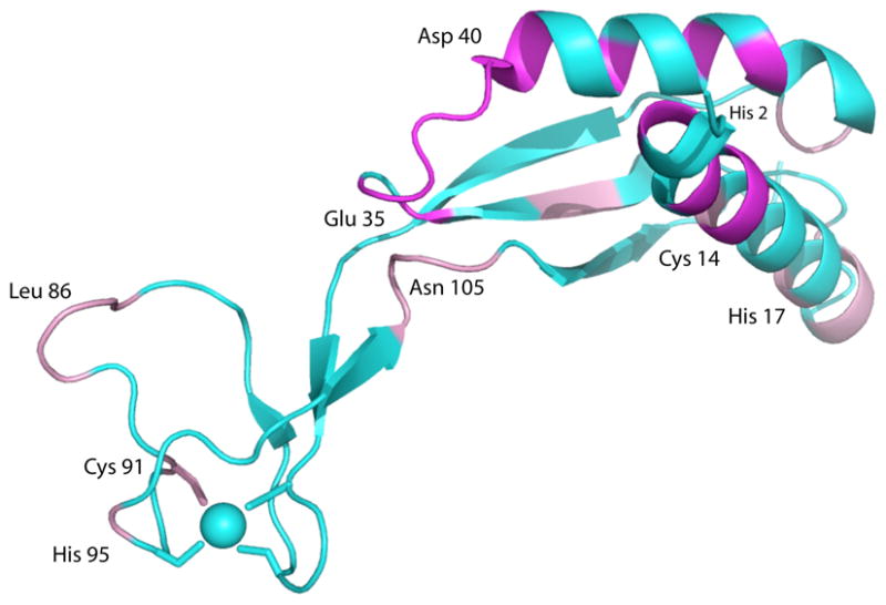
Figure 4.

NMR results superimposed on the structure of monomeric HypA (PDB entry 2KDX).18 Resonances that are broadened to invisibility by addition of Ni(II) ions are shown in magenta. Resonances that are perturbed or doubled by binding Ni(II), indicating more than one conformation is present, are shown in pink. The Zn site is indicated by the blue sphere. See text for details.
