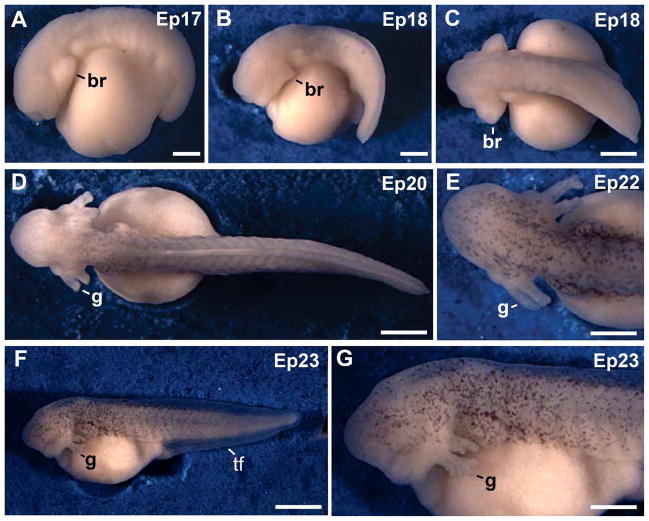
Fig. 2.
External views of embryos from tail bud to hatching. A: Tail bud stage. B: Embryo at the muscular response stage. C: Dorsal view of the embryo in B. D: Embryo at the circulation to the external gills stage. The trunk acquired some dark pigment E: Head region of a stage 22 embryo. Dark pigment was detected in the head region. F: Embryo at hatching. G: Higher magnification of the head region from the embryo in F. The external gills reached their maximum length. Abbreviations: br, branchial arch; g, gills; tf, tail fin. Scale bars: 300 μm in A; 400 μm in E; 500 μm in G; 600 μm B–D; 1 mm in F.

