
Figure 1.
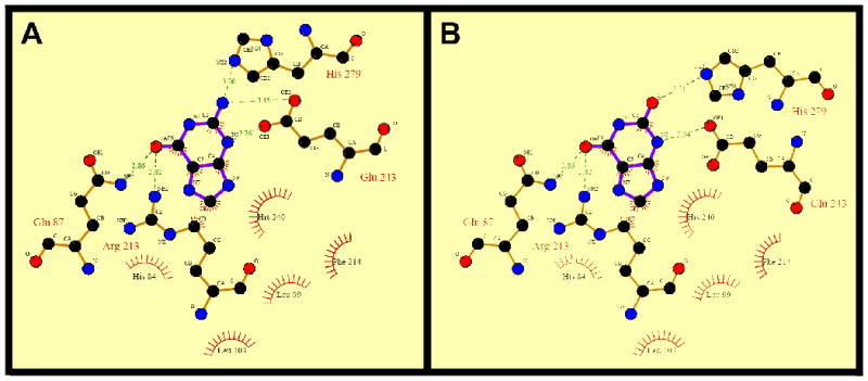
Protein-ligand interactions in the substrate-binding site of human GDA. (A) Guanine-hGDA interactions. (B) Xanthine-hGDA interactions. Two-dimensional representation of ligand–protein interactions were analyzed using Ligplot [54]. Hydrogen bonds formed between protein residues and ligands are indicated by dashed green lines and the corresponding distance (Å) and residues engaged in hydrophobic interactions are represented in red.