Fig. 1.
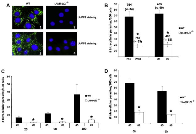
(A) Evaluation of LAMP1 and 2 presence in WT and LAMP1/2−/− cells, through immunofluorescence with anti-LAMP1 (1 and 2) or anti-LAMP-2 (3 and 4) antibodies and secondaries labelled with Alexa Fluor 488®. Cell nucleus is labelled with DAPI. LAMP staining, is observed only in WT cells, while cell nuclei can be seen in all panels. (B) Quantitative analyses of parasite infection in two pairs of Wild type (PS1 and #5) and LAMP1/2−/− (SV48 and #9) fibroblasts. Fibroblasts monolayers were exposed to Tissue Culture derived Trypomastigotes (TCT) from Y strain at an MOI of 50, and then analyzed by immunofluorescence staining. Infection was determined by the number of internalized parasites per 100 counted cells. The total number of fibroblasts per 15 counted fields (with the standard deviation values) is shown on the top of each bar on the graph. (C) Quantitative analysis of parasite infection in #5 (WT) and #9 (LAMP1/2−/−) fibroblasts using different MOI of T. cruzi Y strain (25; 50; 100). Infection was determined as described above. (D) Quantitative analyses of parasite infection in #5 (WT) and #9 (LAMP1/2−/−) fibroblasts, right after or 1 h post-invasion. In this case invasion assays were performed as described in material and methods, however after parasite wash cells were either fixed or re-incubated in media for an additional hour before fixation. Infection was determined as described above. The data in B–D correspond to the mean of triplicates ±SD. Asterisks indicate statistically significant differences (P < 0.05, Student’s t test) between WT and LAMP1/2−/− cells, under the same conditions.

