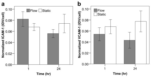
Figure 6.
Quantification of ICAM-1 (a) and VCAM-1 (b) expression on module surface through confocal microscopy and image analysis. The integrated density value (IDV) for ICAM-1 and VCAM-1 was normalized to the total number of visualized cells. No statistically significant differences in expression were found for ICAM-1 or VCAM-1 over the 24 hour time course. Though not statistically significant, ICAM-1 appeared to show a slight upregulation with flow after 1 hour followed by a downregulation after 24 hours (a). VCAM-1 results also suggested a marginal downregulaion with flow at both time points (b). Collectively, this data appears to verify our initial hypothesis that, unlike atherosclerotic-like disturbed flow, perfusion through the tortuous channels of the module bed does not significantly increase the amount of endothelial cell activation. Error bars are ± s.e.m. and n = 6, 7 or 8.

