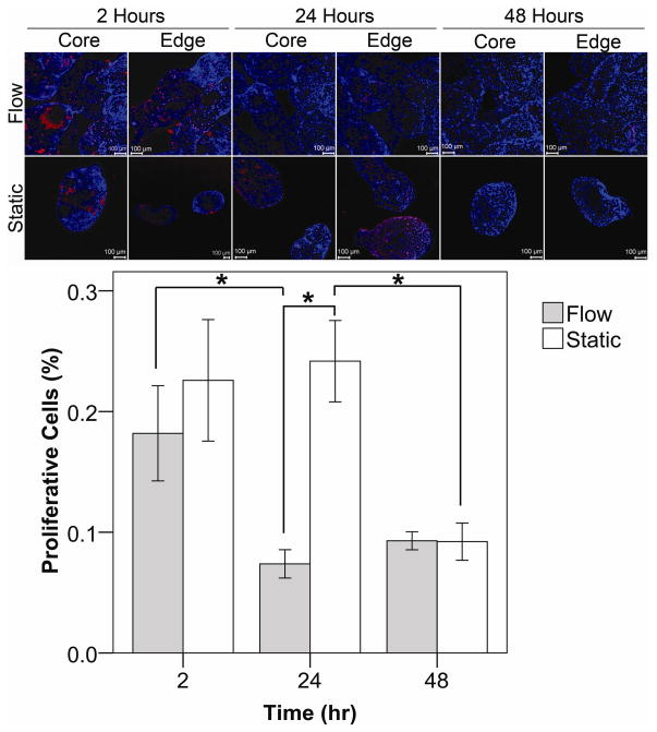
Figure 7.
Collagen modules with a confluent layer of HUVEC were subjected to flow (0.5 mL min−1) for up to 2 days. Proliferating HUVEC incorporated BrdU into their DNA and appeared red. Nuclei were counterstained in blue. The entire packed module bed was examined for proliferation including the interior (core) and perimeter (edge), as indicated. After 2 hours, HUVEC proliferation was similar for both the flow and static cases. After 24 hours of flow, there was less proliferation present, as compared to static controls (p < 0.001) and the 2 hour flow case (p = 0.010). At the 48 hour mark, proliferation for both the static and flow cases became similar as the amount of proliferation in the static controls dropped from its 24 hour high (p <0.05). Error bars are ± s.e.m.,* indicates p < 0.05 and n ranges between 5 and 14. Scale bars = 100 μm.

