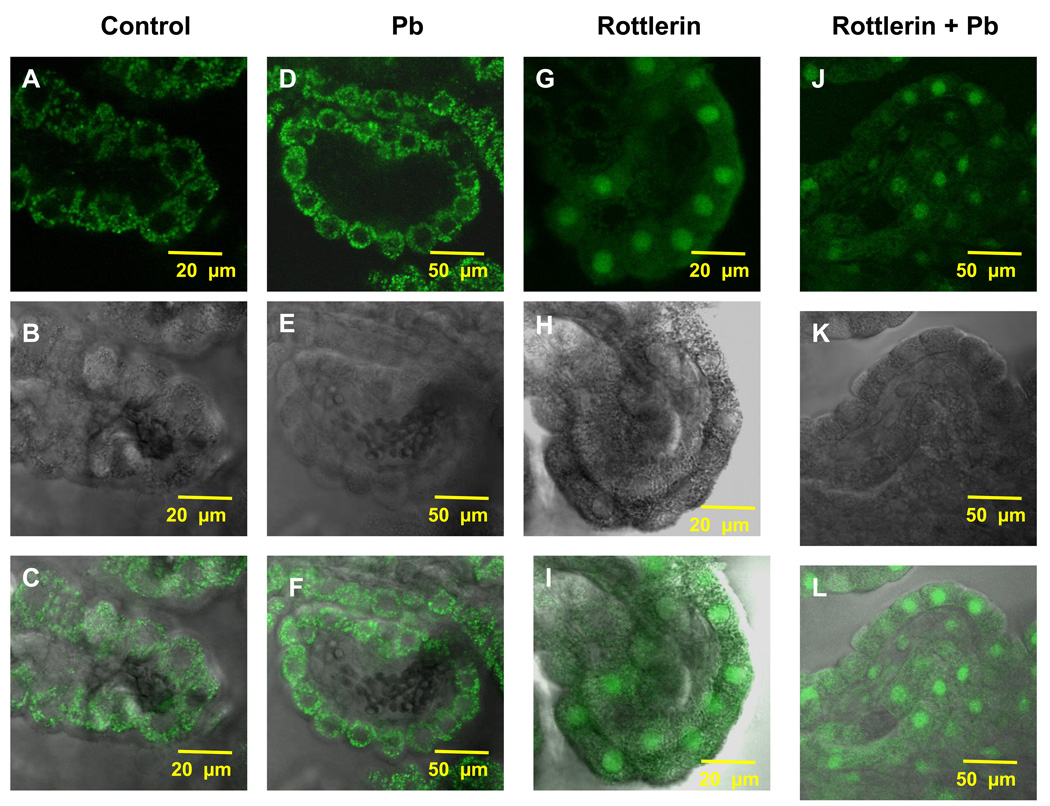
Fig. 5.
Rottlerin attenuated the Pb-induced increase in Aβ accumulation. (A,C) Confocal image from a representative CP tissue of a control rat demonstrating the accumulation of Aβ primarily in the cytosol with minimal staining in the nuclei. (D, F). Confocal image from a representative CP tissue in a Pb-exposed rat demonstrating an increase in Aβ signal. (G, I) Confocal image from a representative CP tissue pre-treated with rottlerin in absence of Pb. (J, L) Confocal image from a representative CP tissue pre-treated with rottlerin in presence of Pb. (B, E, H, K) represent transmission images revealing normal morphology of the tissues. Please see the legend to Fig.3 for Pb-dose regimen. The data are representative of experiments from four replicates.

