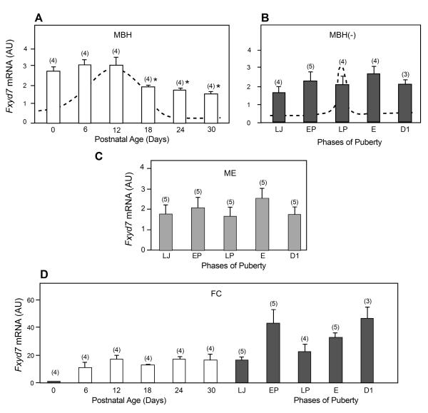
Figure 2.
Pre- and peripubertal changes in Fxyd7 mRNA abundance in the medial basal hypothalamus (MBH), median eminence (ME) and frontal cortex (FC) of female rats, as assessed by real-time PCR. A, The MBH between PN day 0 (neonatal period) and 30 (late juvenile period), B, The MBH, without the ME (MBH-), at the time of puberty, C, The ME alone, D, The FC from PN day 0 to the end of the peripubertal phase, i.e. the day of first dioestrous. Bars are means and vertical lines are SEM. Numbers on top of bars are number of animals per group. For abbreviations see legend to Figure 1. In A,* = p<0.05 vs. PN day 12.

