
Figure 3. CD36 receptor levels and proliferation/migration of liver and lung endothelial cells in vitro.
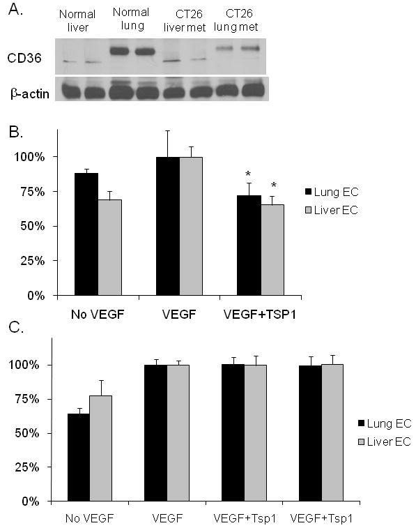
(A) Western blot analysis of CD36 levels in normal mouse liver and lung and CT26 liver and lung metastases. (B) Endothelial cell migration of liver sinusoidal endothelial cells (Liver EC) and lung microvascular endothelial cells (Lung EC) toward VEGF in a modified Boyden chamber with and without TSP1. *p<0.05 compared to VEGF alone. (C) Proliferation of Liver EC and Lung EC with and without VEGF and TSP1.