
Figure 5. Effect of ADAMTS1 knockdown on TSP1 cleavage.
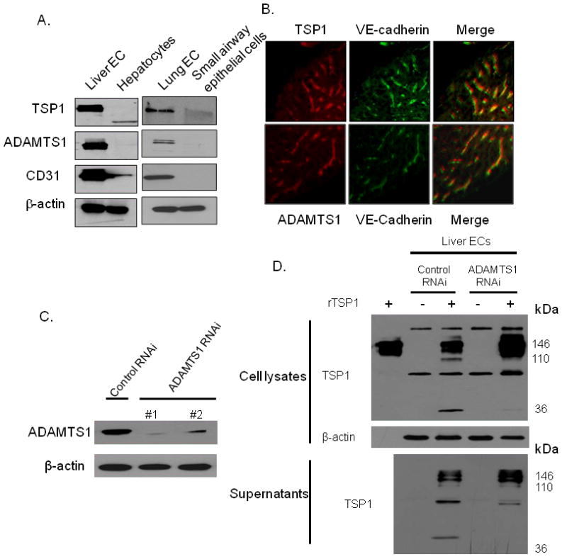
(A) Western blot of TSP1, ADAMTS1, CD31, and β-actin in human liver sinusoidal endothelial cells (Liver EC), hepatocytes, human lung microvascular endothelial cells (Lung EC), and small airway epithelial cells. (B) Co-immunofluorescence for TSP1 (red), ADAMTS1 (red), and/or VE-cadherin in mouse liver tissue. (C) Western blot of ADAMTS1 following infection with ADAMTS1 RNAi lentiviral vectors and control vector. (D) Western blot of TSP1 of Liver EC lysates (upper) and supernatants (bottom) following infection with control lentiviral vector and ADAMTS1 lentiviral vector #1. Lysates or supernatant were incubated with or without recombinant TSP1 protein (rTSP1). Left lane (upper) loaded with rTSP1 alone.