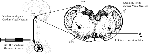
Fig. 1.
Schematic drawing of the experimental preparation. Cardiac vagal neurons (CVNs) were identified by retrograde fluorescent labeling. Slices of 400-μm thickness were obtained and individual identified. CVNs in the nucleus ambiguus (NA) were studied using whole cell patch-clamp technique. The location of the lateral paragigantocellular nucleus (LPGi) was identified using stereotaxic coordinates in addition to the location relative to fluorescently identified CVNs in the NA. Square wave current injections of 0.1–0.5 mA and 1-ms duration were applied to evoke GABAergic pathways from the LPGi to CVNs.

