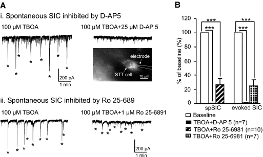
Fig. 6.
SICs are inhibited by blocking and N-methyl-d-aspartate (NMDA) or NR2B receptors. Ai: spontaneous SICs induced by 100 μM TBOA (left) were completely inhibited by bath perfusion (right) of 25 μM d-AP5 in an spinothalamic tract (STT) neuron. The inset shows the STT neuron labeled by a retrograde fluorescent marker. Aii: spontaneous SICs induced by 100 μM TBOA (left) were strongly inhibited by bath perfusion (right) of 1 μM Ro 25–6981 in an unidentified neuron. B: mean (±SE) percent inhibition of the amplitudes of the spontaneous and stimulation-evoked SICs by 25 μM d-AP5 and 1 μM Ro 25–6981. ***P < 0.001.

