FIG. 4.
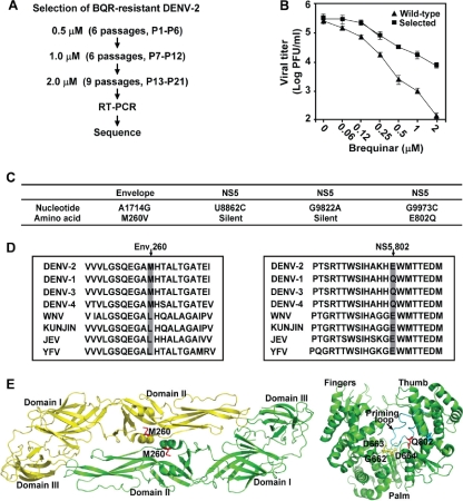
Selection and sequencing of BQR-resistant DENV-2. (A) Scheme for selection of BQR-resistant DENV-2. P1 through P6 were selected at 0.5 μM BQR, P7 through P12 were selected at 1.0 μM, and P13 through P21 were selected at 2.0 μM. (B) Resistance analysis. Vero cells were infected with wild-type or P21 viruses (MOI, 0.1) in the presence of BQR or 0.9% DMSO (as a negative control). At 48 h p.i., the viral titers in culture fluids were quantified by plaque assays. Resistance is quantified by comparison of the viral titers from the BQR-treated infections with the viral titers from the DMSO-treated infections. Error bars indicate the standard deviations from three independent experiments. (C) Mutations recovered from the P21 resistant virus. The locations of the nucleotide and amino acid changes are indicated. (D) Amino acid sequence alignment. (Left panel) Sequence alignment of a flavivirus envelope protein region; (right panel) sequence alignment of a flavivirus RdRp region. The sequences of DENV-1, DENV-2, DENV-3, DENV-4, WNV, KUNV, JEV, and YFV are derived from the sequences with GenBank accession numbers U88535, M29095, M93130, AY947539, AF404756, D00246, AF315119, and X03700, respectively. (E) Locations of resistance mutations on crystal structures of DENV-2 envelope protein and DENV-3 RdRp. (Left panel) DENV-2 envelope protein structure (Protein Data Bank code 1OKE [20]). The two envelope protein subunits are in yellow and green; the mutated residue M260 is labeled in red. (Right panel) DENV-3 RdRp structure (Protein Data Bank code 2HFZ [36]). The fingers, palm, and thumb subdomains are indicated; the GDD active site of RdRp is shown in yellow; the priming loop is colored in cyan; the mutated residue Q802 is shown in red. The images were prepared using the PyMol program.

