Figure 1.
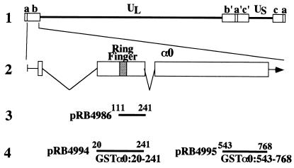
(A) Schematic diagram of the sequence of the HSV-1 genome and the location of the α0 gene. Line 1, a linear representation of the HSV-1 genome. The unique sequences are represented as the regions. The terminal repeats flanking the unique long (UL) and unique short (US) sequences are shown as open rectangles with their designation letters shown above. Line 2, an expanded section of the domain encoding α0 gene. The transcribed and coding domains are shown for one copy of the α0 gene. An identical copy is located in the inverted repeat (b′) flanking UL. Line 3, the region used as “bait” in the yeast two-hybrid screen. Line 4, the domains of the α0 gene fused to GST to generate chimeric proteins.

