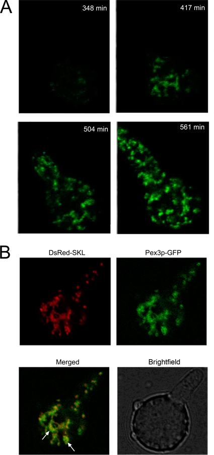
FIG. 5.
Peroxisomes form from a network in germinating P. chrysogenum conidiospores. (A) Stills from Video S1 in the supplemental material at various time points during conidiospore germination. The first fluorescence is observed as a relatively small network structure at approximately 7 h of incubation of the spores in fresh PEN production medium. This structure rapidly increases in size at later stages of incubation and extends into the germination tube. (B) CLSM pictures of germinating conidiospores that simultaneously produce DsRed-SKL and Pex3-GFP. The merged image shows that the two fluorescent probes colocalize, indicating the peroxisomal nature of the complex network. Arrows point to the reticular networks that contain both DsRed-SKL and Pex3-GFP.

