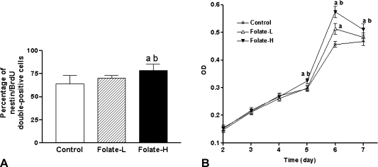
Fig. 2.
Folic acid supplementation stimulates proliferation of embryonic neural stem cells (NSCs). A: Percentage of cells that were double-positive for nestin and 5'bromo-2'deoxyuridine (BrdU), as determined by immunofluorescence analysis. B: Cell proliferation quantified by measuring the time-dependent increase in the abundance of viable cells, which were identified by methyl thiazolyl tetrazolium (MTT) assay. NSC neurospheres were incubated at 37°C, for the indicated periods, in medium containing 4 mg/l folic acid (Control), 8 mg/l folic acid (Folate-L), or 44 mg/l folic acid (Folate-H). MTT was added to each well at 4 h before the end of the incubation. Finally the resulting precipitates were dissolved in dimethyl sulphoxide and optical density (OD) was measured at 490 nm. Shown are mean ± SD values of 8 separate experiments. One-way analysis of variance and Tukey’s multiple comparison were performed to compare the mean values. ap<0.05 vs Control, bp<0.05 vs Folate-L.

