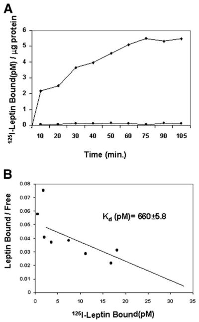
Fig. 3.
Specific binding of 125I-leptin to HSCs. (A) Time course showing leptin binding to HSCs. Cells were cultured as monolayers and incubated with 62.5 pmol/L 125I-leptin for time indicated in minutes (◆). To determine specific leptin-HSC binding, an identical assay was performed with excess unlabeled leptin (5 μmol/L) (●). Bound radioactive ligand was measured and standardized to microgram protein assayed.28 (B) Scatchard analysis.29 Incubation with labeled leptin was performed for 2 hours with cell monolayers at 4°C. Cell-associated radioactivity was determined by scintillation counting. Data for Kd are expressed as mean ± SEM. The plots represent duplicate experiments performed twice. The details of these studies are described in Materials and Methods.

