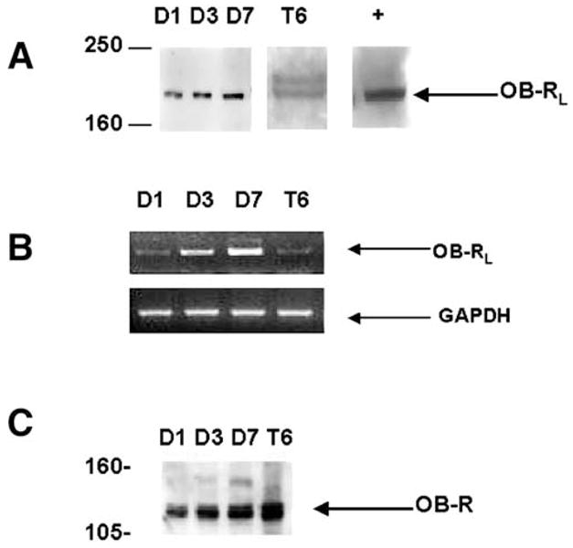
Fig. 4.

Detection of OB-RL in HSC-T6 cells and primary, culture-activated rat HSCs by immunoprecipitation and RT-PCR. (A) Immunoprecipitation of OB-RL. Whole-cell lysate was prepared from primary HSCs in culture for 1 day (D1), 3 days (D3), and 7 days (D7). OB-RL in HSC-T6 (T6) cells was detected by immunoprecipitation. An equal quantity of protein was immunoprecipitated with antibody against the cytoplasmic domain of the leptin receptor. Positive control was from mouse brain (+) and was supplied by Santa Cruz Biotechnology. (B) RT-PCR detecting mRNA for OB-RL. Details of reactions and primer pairs are described in Materials and Methods. Total RNA was isolated from primary HSCs and HSC-T6 in culture at times identical to immunoprecipitation analysis as previously described. The expression of glyceraldehyde-3-phosphate dehydrogenase (GAPDH) was used as an internal control. (C) Immunoprecipitation of OB-R with antibody directed at the extracellular domain of the receptor common to all OB-R isoforms. Molecular weight is on the left in kilodaltons.
