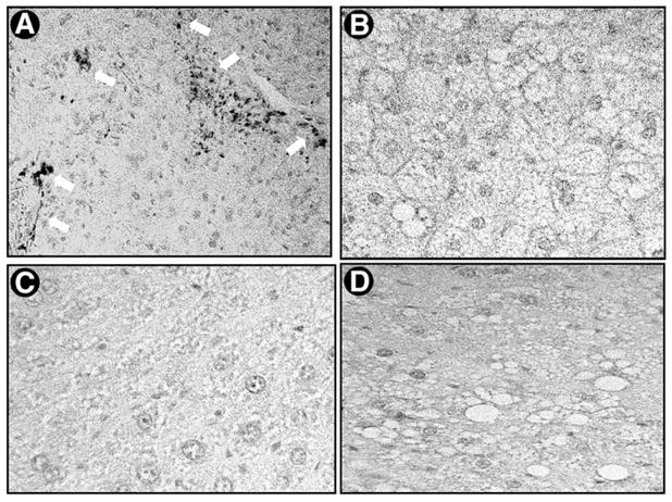
Fig. 7.

Expression of α-SMA in the liver after administration of CCl4 in ob/ob mice and their lean littermates. The expression and localization of α-SMA were detected by immunohistochemical staining as described in detail in Materials and Methods. Representative micrographs from 3 individual experiments are shown (original magnification 400×). (A) CCl4-treated lean littermates showing α-SMA–positive cells (white arrows), (B) CCl4-treated ob/ob mice, (C) vehicle-treated lean littermates, and (D) vehicle-treated ob/ob mice. Primary antibody dilution was 1:400; the experimental design and resultant liver sections used for α-SMA staining were as in Fig. 6.
