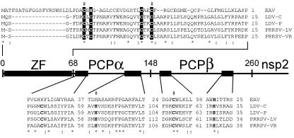
Figure 5.
Identification of a ZF motif in the N-terminal domain of the arterivirus replicase. The sequences of lactate dehydrogenase elevating virus [LDV-C (44) and LDV-P (45)], porcine reproductive and respiratory syndrome virus [PRRSV-LV (46) and PRRSV-VR2332 (47)], and EAV (6) were compared. Alignments of the complete ZF domain and selected conserved regions of PCPα and PCPβ containing the active-site Cys and His residues (bold) are shown. Invariant (*) and conserved (:) positions are highlighted. Conserved His and Cys residues in the nsp1 ZF that are proposed to be involved in zinc binding are shown in reverse shading, and mutated residues (Table 1) are indicated with “#.”

